Digital tools are transforming diagnosis, design, and restoration from manual processes into data-driven workflows—faster, more accurate, and more comfortable for patients.
Digital dental solutions refer to an integrated approach that connects scanning, imaging, design, manufacturing, and management through digital tools. Core components include intraoral scanners, laboratory scanners, CAD (design) software, CAM (milling/printing) equipment, 3D printers, sintering/curing devices, along with compatible materials and cloud/local data management systems. It is not a single device but a traceable, repeatable, and measurable workflow.
Higher Precision, Fewer Re-works: Numerous systematic reviews demonstrate that digital impressions achieve equivalent or superior spatial accuracy compared to traditional impressions across multiple clinical scenarios, particularly excelling in single crowns, small-span bridges, and certain implant-guided applications.
Significantly Improved Time Efficiency: The total cycle time from impression taking, design, fabrication to installation is substantially reduced; this is especially evident in practices offering chairside same-day restorations.
Enhanced Patient Experience: Eliminates uncomfortable impression materials and enables more intuitive communication (e.g., displaying fit simulations on screen).
Cost control (long-term): Initial investment in equipment and consumables is higher, but reduced rework, shorter labor hours, and faster turnaround yield long-term returns.
Traceability and data management: Digital records facilitate quality control, remote collaboration, and regulatory compliance (as required by region).
Rapid market and technological growth: The overall digital dentistry market is expanding, with the industry ecosystem and supporting services evolving.
Grand View Research
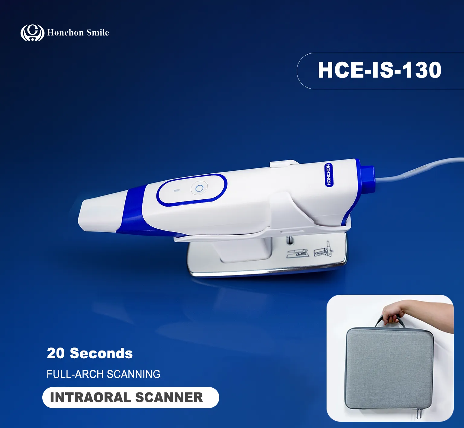
Purpose: Replaces traditional impressions to generate digital files (STL/PLY, etc.).
Selection Focus: Clinical accuracy, scanning speed, seam-smoothing (stitching algorithm), tolerance for wet/reflective areas, open/closed system, software ecosystem, and after-sales support.
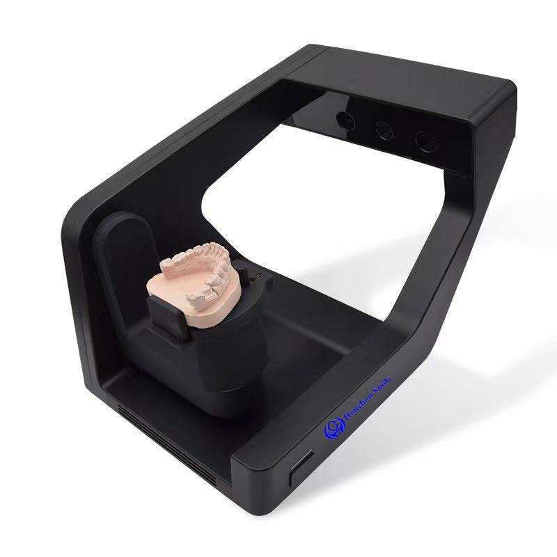
Purpose: Scans plaster models, articulators, or impressions for laboratory design and fabrication.
Selection Focus: Resolution, repeatability, interface compatibility.
CAD Software
Function: Restorative design, occlusal adjustment, parametric templates, automated design plugins (e.g., implant guidance, immediate implant placement).
Selection Focus: Compatibility with existing scanners and CAM systems, learning curve, update and support policies.
CAM: Milling Machines & Sintering Furnaces
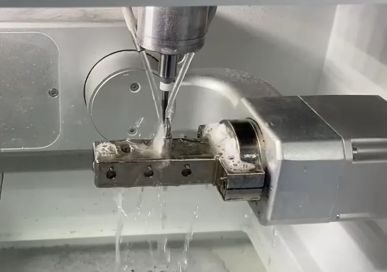
Milling: Dry/wet milling, axis count (3/4/5-axis), tooling ecosystem, compatible materials (zirconia, glass ceramics, resins, etc.).
Sintering Furnaces: Sintering curve stability, material library, energy consumption, and lifespan.
3D Printers and Materials
Applications: Models, guides, sleeves, orthodontic attachments, guided implants, and some for final restorations (depending on material regulations). Dental 3D printing adoption and market are rapidly expanding.
Grand View Research
Key Considerations: Print accuracy, repeatability, material types (resins, biocompatible materials), post-processing workflows (cleaning, curing), safety, and regulatory compliance.
AI and Imaging-Assisted Tools
Trend: AI is being applied to image diagnosis (aiding X-ray and CBCT interpretation), case screening, caries/periodontal risk assessment, and workflow automation, transitioning from research to clinical tools. Consider explainability, validated studies, and privacy compliance during selection.
Frontiers
In-Office Scanning / Image Acquisition (IOS, CBCT)
File Transfer and Inspection (STL/PLY/OBJ; check for missing data, alignment errors)
Design (CAD): Set boundaries, occlusion, gaps, and cutting allowances.
Simulation and Virtual Try-in (Optional): Aesthetic evaluation, occlusal simulation.
Manufacturing: Select milling or printing, configure tooling/print parameters.
Post-Processing: Support removal, polishing, staining, sintering/curing.
Clinical Try-In and Adjustment: Fine-tuning, final retention.
Archiving and Feedback: Save digital records, document issues for workflow optimization.
Practical Tip: Pre-set standard checklists in CAD (clearance, wall thickness, bridge span, inlay orientation) to detect and correct common errors early.
Investment Evaluation: Factor in training, consumables, maintenance, software subscriptions, and downtime costs beyond equipment. Recoup partial costs short-term via increased throughput and reduced rework; same-day restoration clinics achieve faster ROI.
Grand View Research
Staff Training: Develop role-specific training plans for scanners, technicians, and clinicians, allowing for trial-and-error periods.
Process Standardization: Establish SOPs covering scanning to restoration (including file naming, version control, quality checkpoints).
Collaboration with External Labs/Cloud Platforms: Select open systems for interoperability with multiple labs to mitigate vendor lock-in risks.
Regulatory & Material Compliance: For clinical use of final restorations, verify materials and processes meet local medical device regulations (CE, FDA, etc.) and sterilization/disinfection requirements.
Scan Misalignment/Misregistration: Causes often include improper scanning angle, saliva, reflective objects, or scanning strategy. Solutions: Optimize scan path, position saliva ejector, use scanning powder (device dependent).
Poor Milling Fit (Poor Interlock): Check CAD-set clearances, tool wear, and fixture positioning.
3D Print Warping or Layering: Review print orientation, support strategy, layer thickness, and post-curing process.
Sintering Shrinkage Management (Zirconia): Use appropriate scaling factors and validated curves, with sample verification.
AI Deep Integration in Diagnosis and Workflow Automation: AI will extend beyond image interpretation to include case screening, automated design suggestions, and quality control, reducing repetitive tasks.
Frontiers
3D Printing as Core Manufacturing Capability: Expanding beyond models to more end-use parts (with material advancements and regulatory approvals), 3D printing will replace certain traditional milling steps.
Grand View Research
Cloud Collaboration and Remote Laboratory Ecosystems: Data interconnectivity, remote design, and remote image review will become standard, facilitating cross-regional services.
Robotic and Automated Surgery/Procedures (under development and testing): Faster automated preparation and repair processes may emerge in the future (still requiring ethical and regulatory validation).
Define Objectives: Is the goal to shorten delivery times, improve quality, or expand same-day restoration services?
Pilot First: Select 1–2 common indications for digital piloting (e.g., single crowns, guides).
Prioritize Equipment Compatibility: Choose open systems and standard file formats (STL/PLY).
Establish Training Records and SOPs.
Implement Quality Checkpoints: Four critical inspections—scanning, design, manufacturing, and try-in.
Track KPIs: Rework rate, delivery cycle, patient satisfaction, unit cost.
Sign clear SLAs (Service Level Agreements) with material/software suppliers.
Expand Gradually: Stabilize processes before adding equipment or launching same-day repairs.
Digital dentistry isn't an overnight “Great Leap Forward.” It's the accumulation of measurable small improvements that collectively yield significant efficiency and quality gains. Adopting the principle of “controlled piloting, standardized implementation, and gradual expansion” minimizes risk and achieves mid-term ROI. Need this adapted into English, condensed into a one-page PowerPoint outline, or customized into an implementation plan tailored to your clinic/lab's specific equipment and personnel? I can directly create a template for your use.